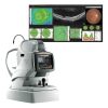
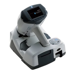
The Retina Scan Duo 2 is a combined OCT and fundus camera system that incorporates new features enhancing screening and clinical efficiency in addition to user-friendly features that were incorporated from the previous model.
FEATURES:
- Fundus image acquisition with macula and disc capture in one image on OCT
- Combined diagnosis of macular and disc pathologies
- Wide area scan (12 x 9 mm)
Wide area normative database (macula: 9 x 9 mm, disc: 6 x 6 mm): A 12 x 9 mm wide area image can be acquired. The retina map captures both the macula and disc in a single shot. The normative database provides a wide area color-coded map comparing the patient’s macular thickness to a population of normal eyes.
- Denoising technique with deep learning: A new image enhancement technique using deep learning automatically displays a denoised image once B-scan acquisition is complete. With deep learning of a large data set of images averaged from 120 images, this denoising technique provides high definition images comparable to a multiple-image-averaging technique. The denoising function generates high definition images from a single frame while decreasing image acquisition time and increasing patient comfort.
- Quick acquisition of high definition B-scan images from a single-frame image
- Fundus autofluorescence (FAF): The FAF function is an advanced screening feature that allows non-invasive evaluation of the RPE without contrast dye.
*Available for the FAF model
Spectification
Nidek RS-330 Retina Scan Duo Specification:
370 (W) x 536 (D) x 602 (H) mm / 39 kg
OCT scanning
Principle spectral domain OCT
Optical resolution optical Z: 7 um, X-Y: 20 um
Scan range: X 3 to 12 mm, Y 3 to 9 mm, Z 2.1 mm
Digital resolution Digital Z: 4um, X-Y: 3um
OCT light source: 880nm
Scan speed: Max. 53,000 A-scan/s (regular mode)
Acquisition time of 3D image: 1.6 s (regular mode)
Auto alignment: Z direction
Minimum pupil diameter: o2.5 mm
Scan patterns: Macula line, macula cross, macula map, macula multi, macula radial, disc circle, disc map, disc radial
Fundus surface imaging
Principle: OCT phase fundus
Angle of view: 40 degree x 30 degree
Fundus camera
Non-mydriatic fundus camera, color, FAF (optional)
Angle of view: 45 degree
Minimum pupil diameter: o4 mm
Light source: Xenon �ash lamp 300 Ws
Flash intensity: 17 levels from F1 (F4.0 +0.8 EV) to F17 (F16 +0.8 EV) 0.25 EV increments
Camera: Built-in 12-megapixel CCD camera
Working distance: 45.7 mm
Display: Tiltable 8.4-inch color LCD
Dioptric compensation for patients eyes :
-33 to +35 D total
-33 to -7 D with minus compensation lens
-12 to +15 D with no compensation lens
+11 to +35 D with plus compensation lens
Internal fi�xation lamp: LED
Horizontal movement: 36 mm (back / forth), 85 mm (right/left)
Vertical movement: 32 mm
Chinrest movement: 62 mm (up/down, motorized)
Auto tracking: +-16 mm (up/down), +-5 mm (right/left), +-5 mm (back/forth)
Document
Nidek RS-330 Retina Scan Duo Brochure Download