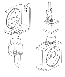


The OCULUS ImageCam® 3
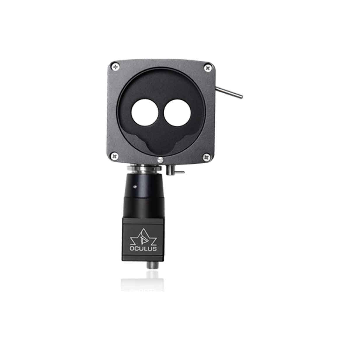
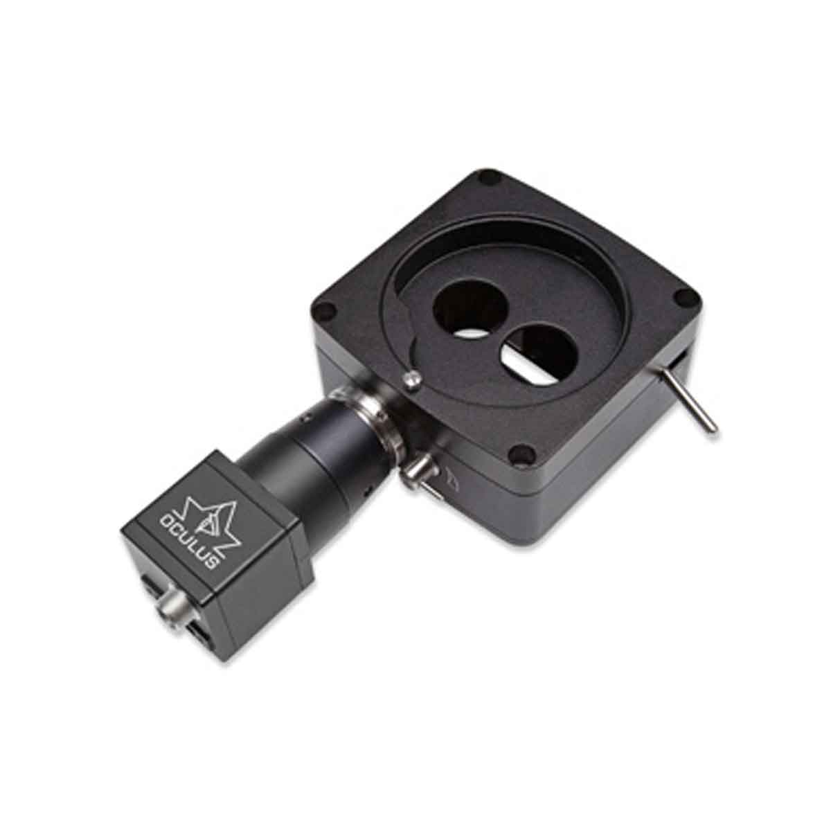
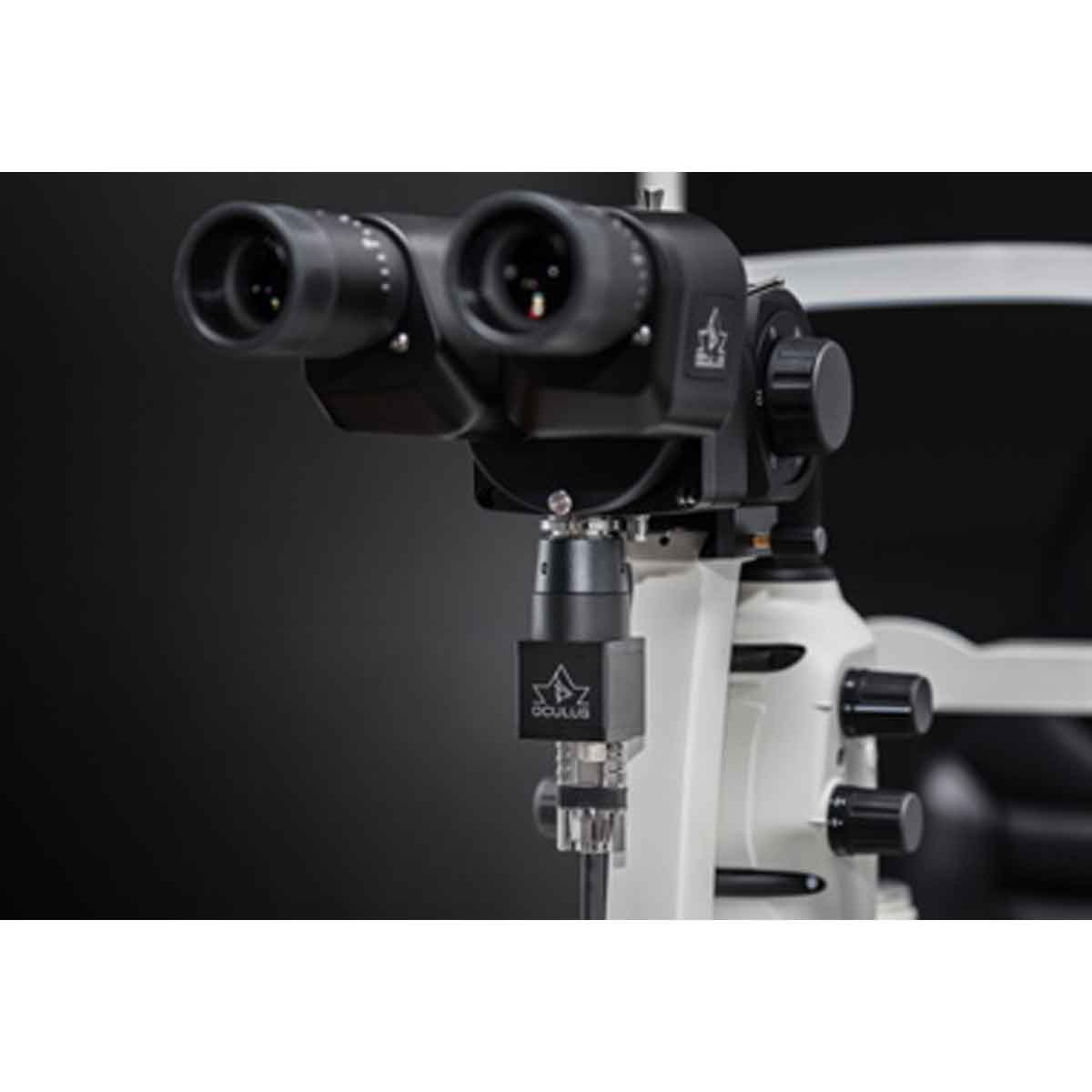
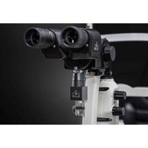
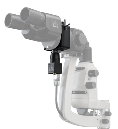
- Small and lightweight – The beam splitter and the camera unit are extremely small and lightweight, making your routine slit-lamp diagnostics light work in the truest sense of the word.
- Swivelling prism – The coupling prism of the ImageCam® 3 beam splitter can be completely swung in and out of the slit lamp’s beam path using a lever.
- Universally adaptable – The OCULUS ImageCam® 3 system is suitable for most slit lamps thanks to its versatile adaptation options.
- Super light sensitive USB 3.0 camera – Only a powerful camera can produce low-noise images in as difficult conditions as prevail around the eye.
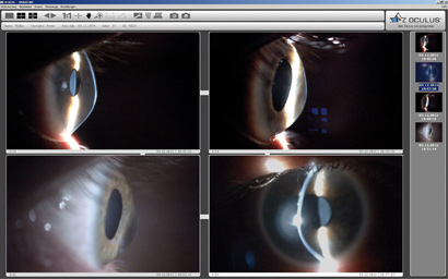
- Intuitive Software – The software is easy to use, presenting the images recorded in an attractive frame.
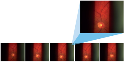
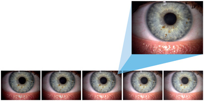
- Image from video – The ImageCam® 3 enables you to record your entire slit lamp examination on a video from which you can then select the best frames for your archive. That spells good-bye to blurred images and consistently high-class images for your documentation.