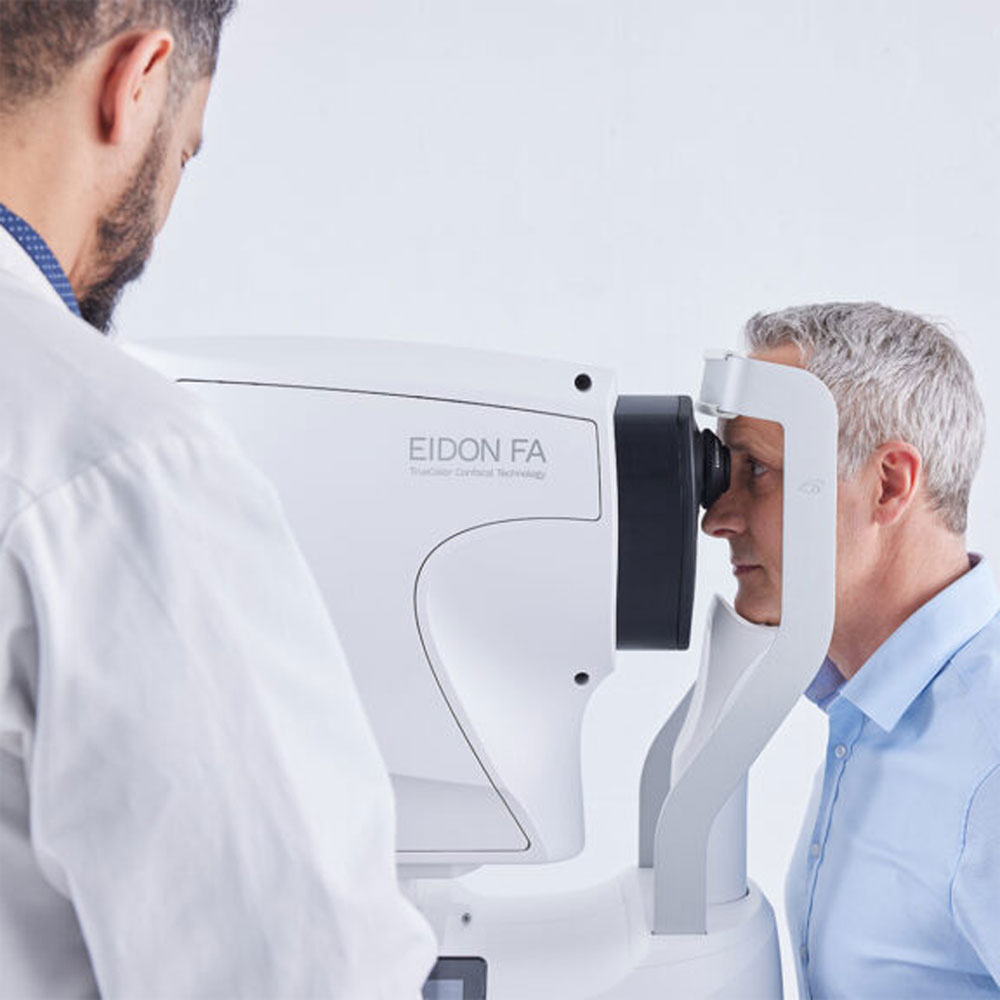
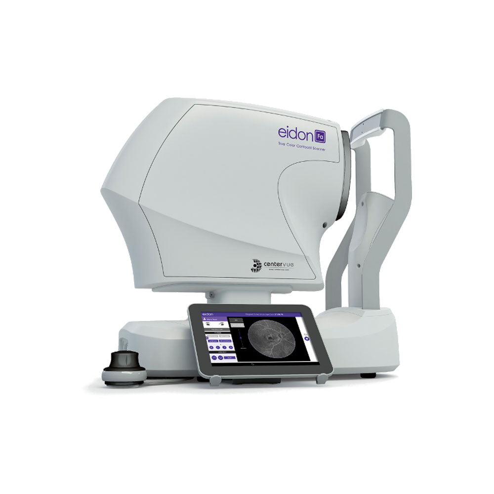
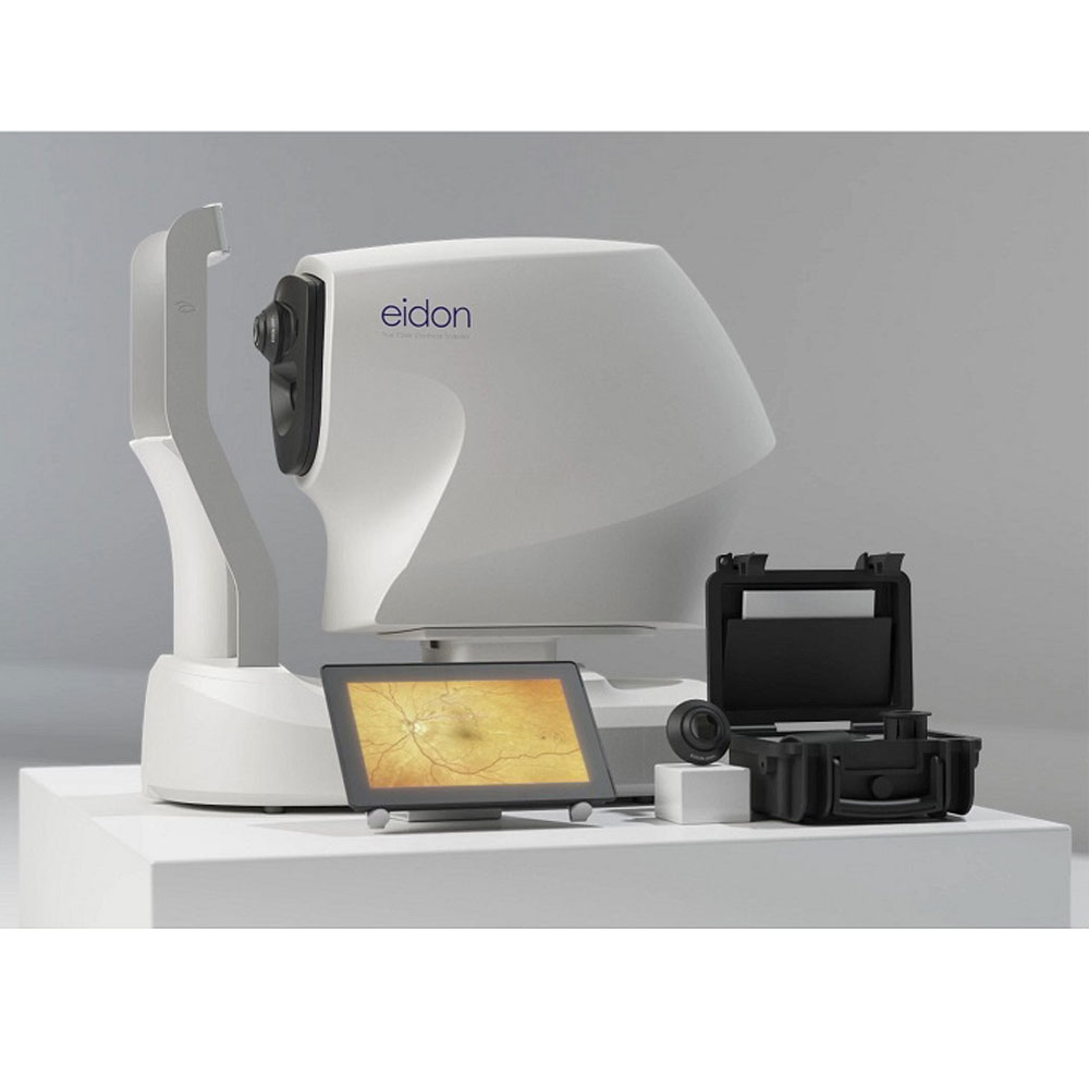
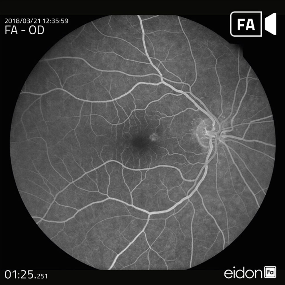
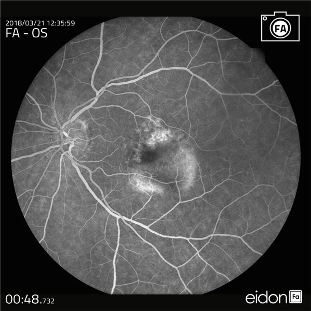
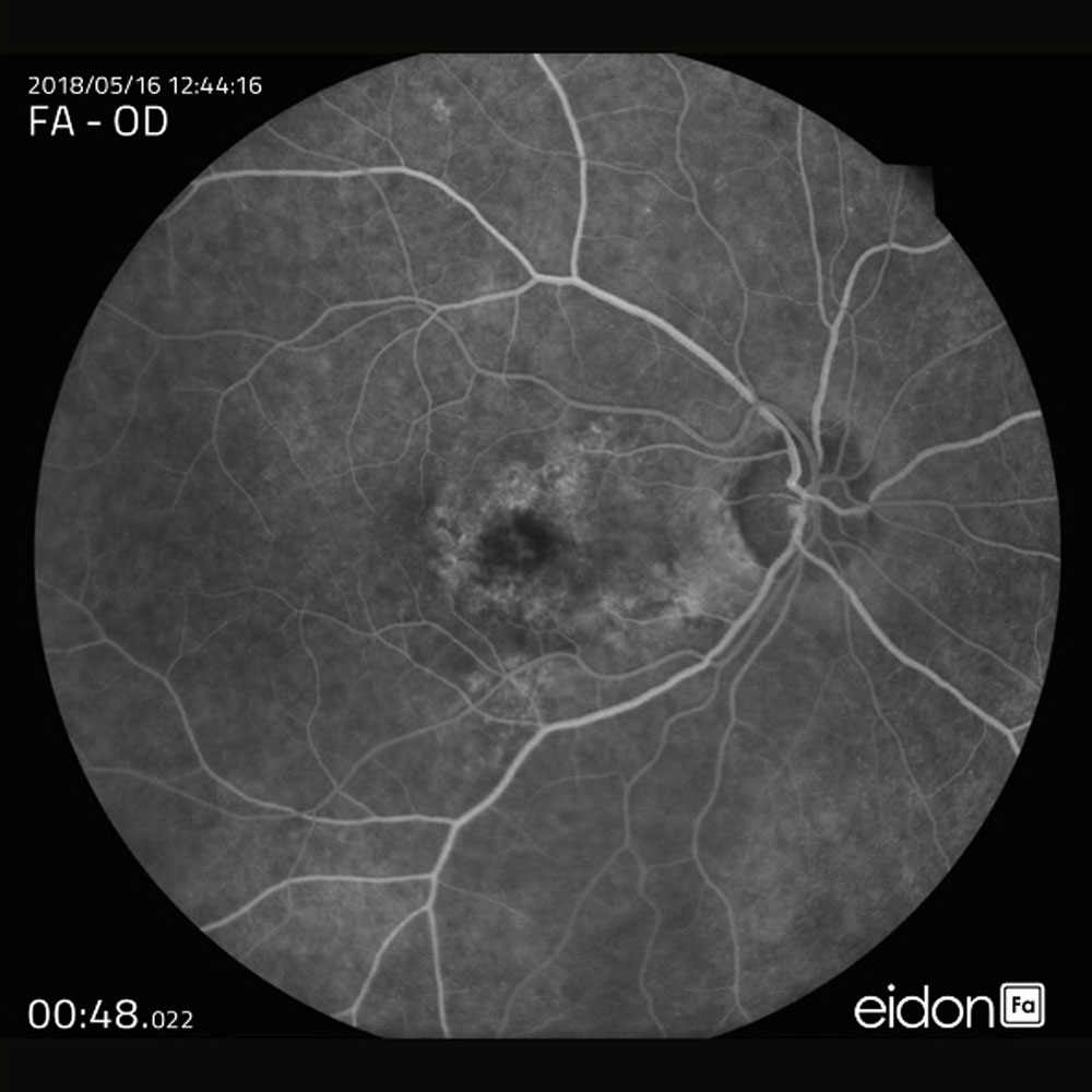
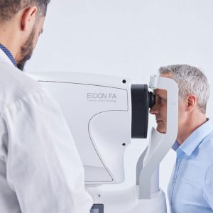
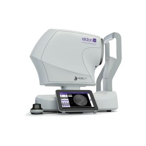
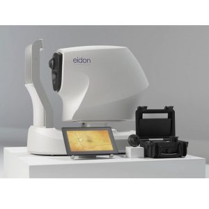
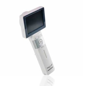
iCare EIDON FA with Fluorescein Angiography capability for ultra-high resolution and Widefield images and videos
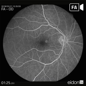
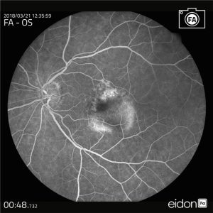
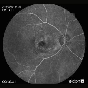
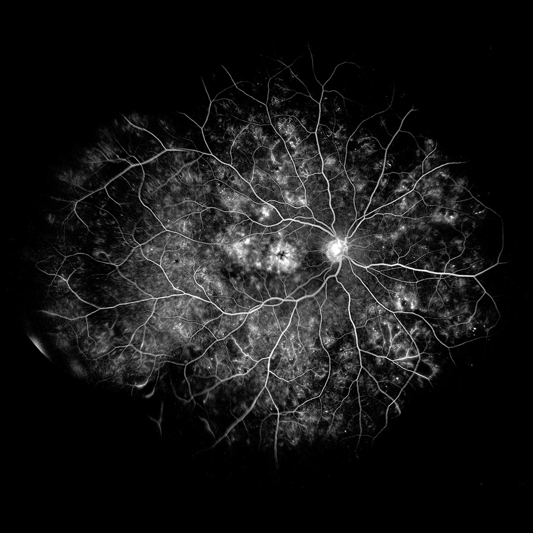
iCare EIDON FA is a top-end imaging system that combines the best of the iCare EIDON technology with automatic Fluorescein Angiography capability to provide a complete suite of imaging modality of unsurpassed image quality. It offers widefield imaging of Fundus Fluorescein Angiography while preserving the image quality, sharpness, and details, even in the periphery.
iCare EIDON FA also offers the added advantage of capturing a detailed ultra-high resolution FA video recording which provides a realistic and dynamic view of retinal vasculature and circulation mechanisms that may be missed with static flash photography.
Fluorescein angiography video for detailed dynamic view
A dynamic perspective offers a realistic representation of retinal vasculature and related circulation mechanisms that may be missed with static flash photography. iCare EIDON FA can capture ultra-high resolution video of the Fluorescein Angiography eliminating the pressure to capture perfect single FA-images when the dye is introduced to the eye.
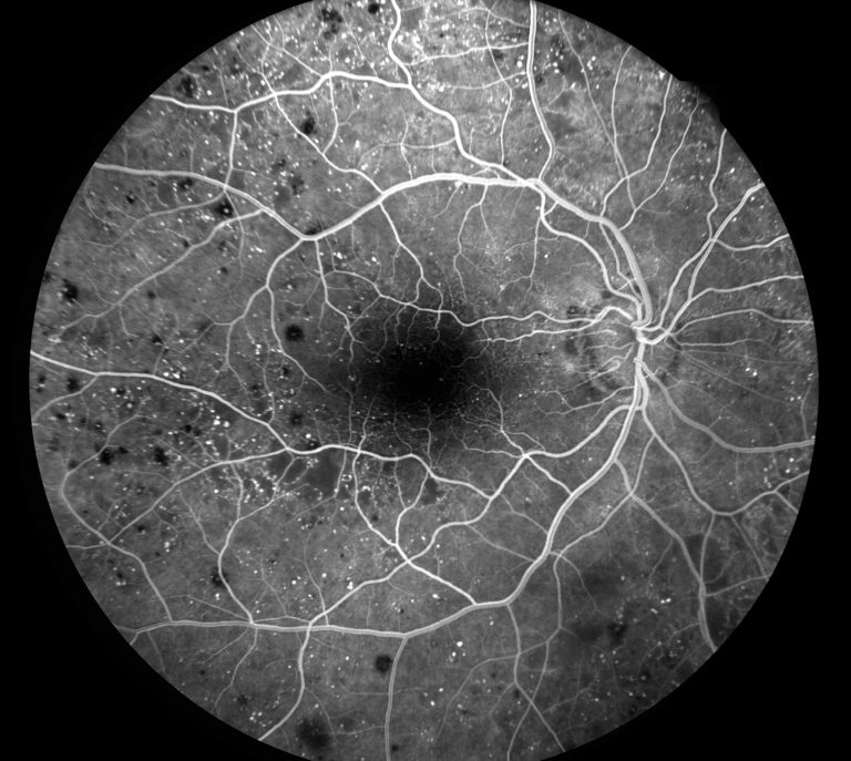
With the advantage of video acquisition capability, the iCare EIDON FA captures a clear, sharp, and detailed dynamic video view accurately and efficiently — leaving the operator free to focus all attention on ensuring maximum patient care and comfort. The video lets you choose the best from multiple frames to accurately document the pathology.
A complete suite of fundus imaging capabilities
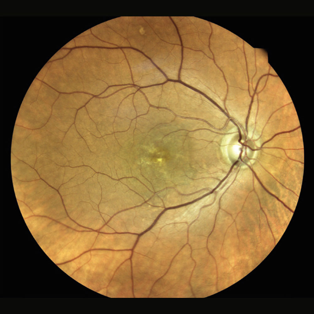
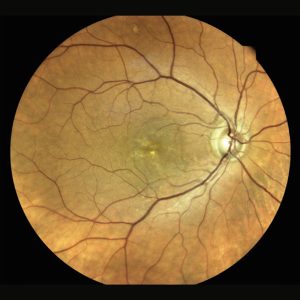
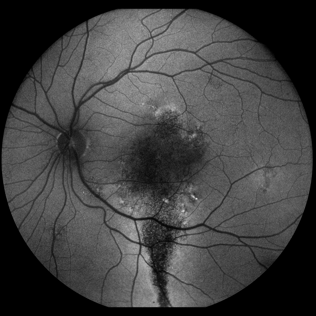
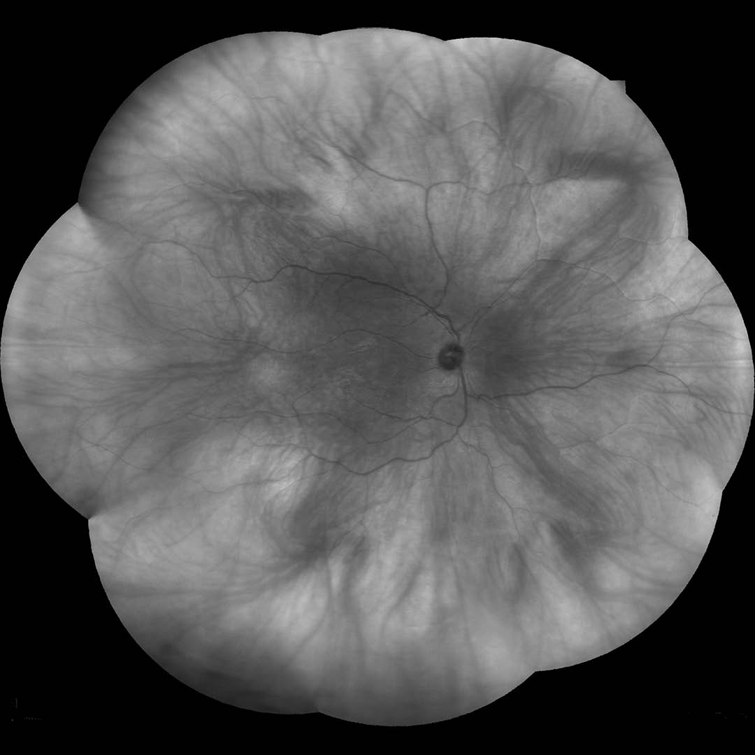
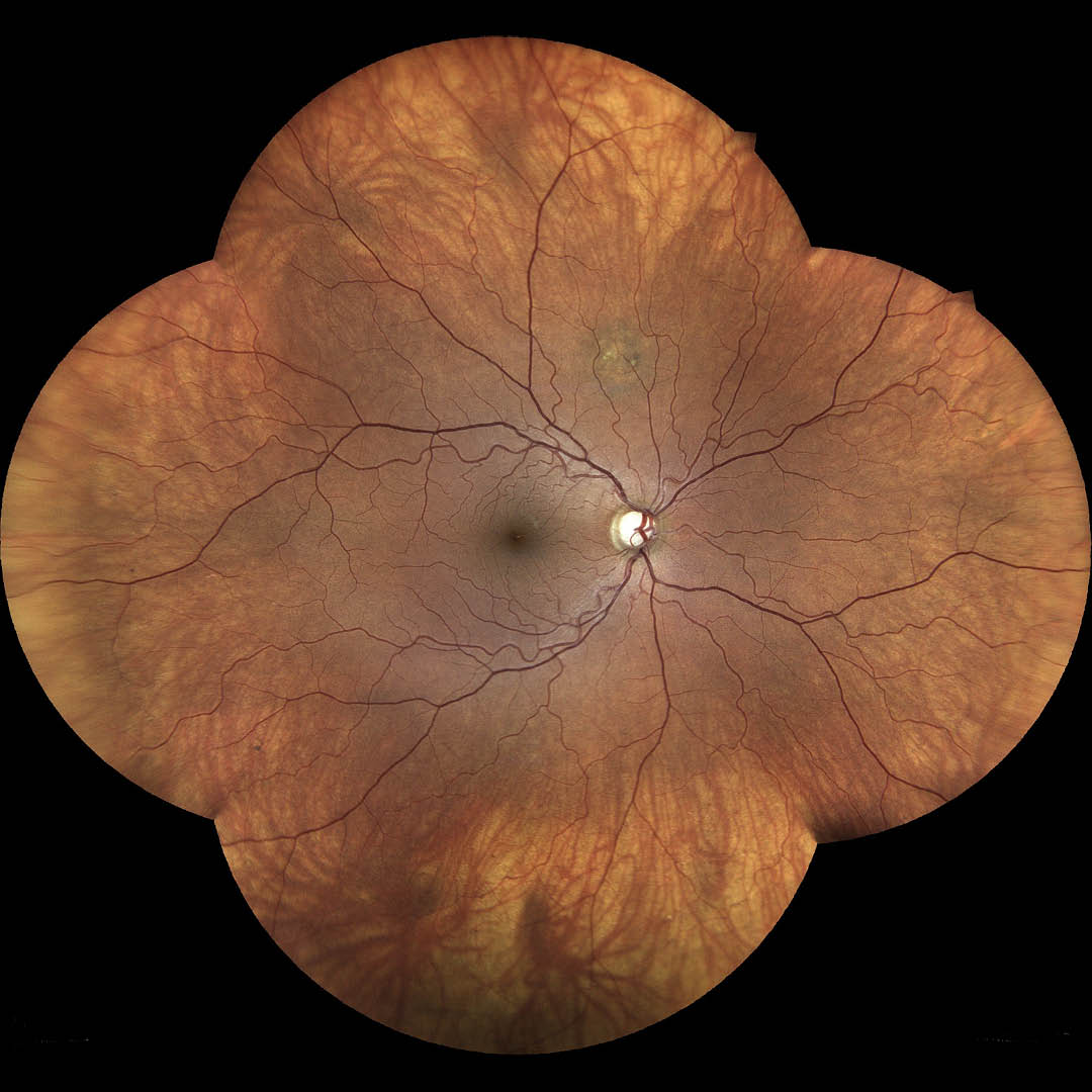
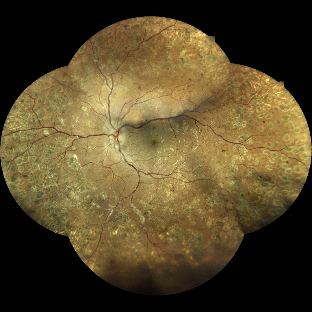
iCare EIDON FA is an extraordinary tool for obtaining multiple types of high value information from multiple imaging modalities. White LED illumination provides high-quality TrueColor images, Red-free filtering enhances visualization of retina vasculature, blue images provide improved view of the Nerve Fiber Layer (RNFL) and red channel allows light to penetrate into the deep layers of the retina. Infrared light provides detailed information corresponding to the choroid. Autofluorescence allows the assessment of the Retinal Pigment Epithelial (RPE) layer integrity. Fluorescein Angiography images and videos enable the clinician to observe and monitor retinal blood flow details.
Increased field of view up to 200˚
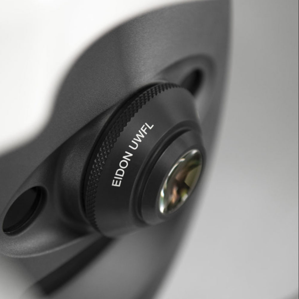
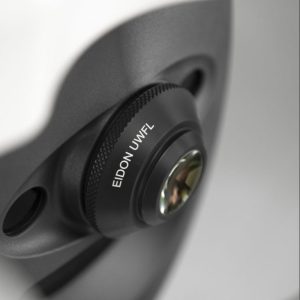
Thanks to the EIDON Ultra-Widefield Module it is possible to increase the field of view up to 200˚, which helps to detect signs of pathologies that start to appear in the periphery. The Ultra-Widefield module enables retina from 120° with a single shot, up to 200° with Mosaic functionality.

